Typical Animal Cell Seen Under Electron Microscope - Electron Microscope Eukaryotic Animal Cell - Micropedia : Most cells, both animal and plant, range in size between 1 and 100 micrometers and are thus visible only with the aid of a microscope.
byEdgar Celuch-
0
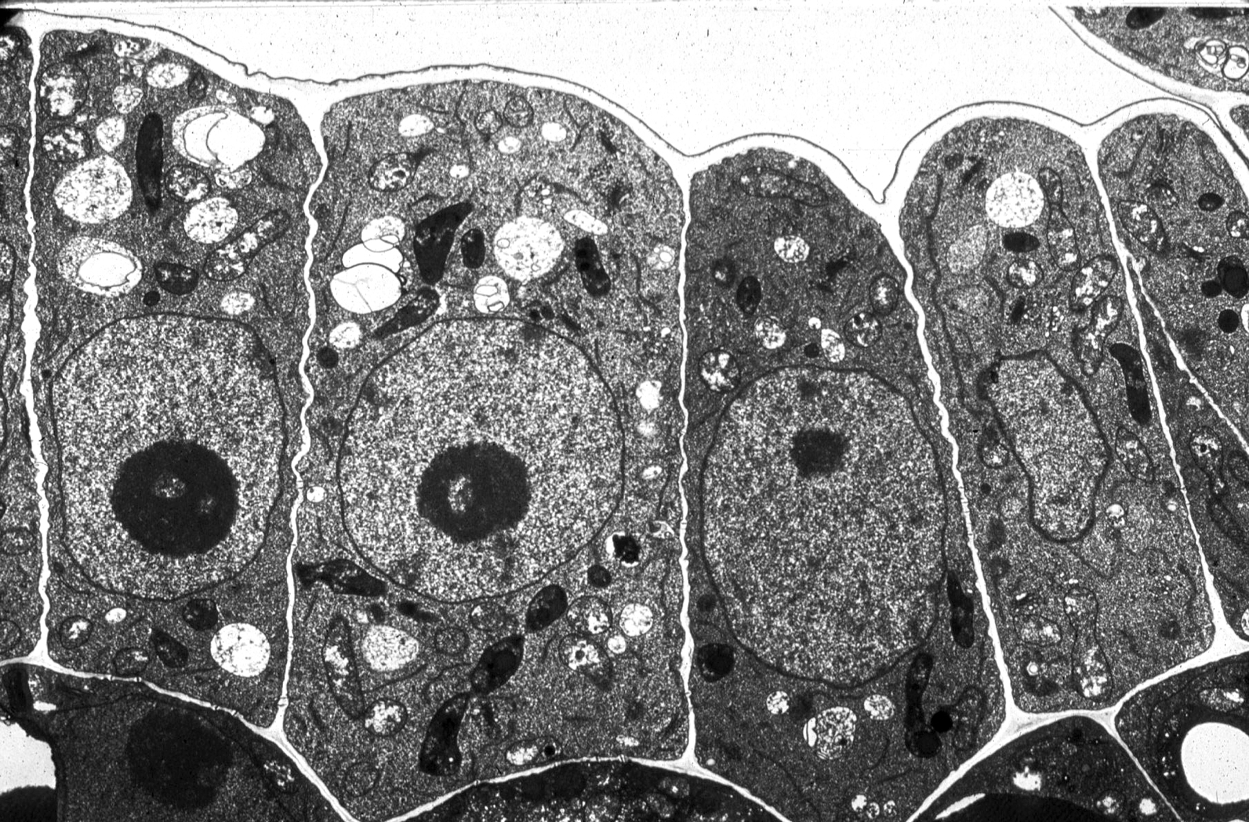
Typical Animal Cell Seen Under Electron Microscope - Electron Microscope Eukaryotic Animal Cell - Micropedia : Most cells, both animal and plant, range in size between 1 and 100 micrometers and are thus visible only with the aid of a microscope.. Animal cells under a microscope. Cristae many infolding that increase surface area to hold more electron carriers; Animal cell under a microscope. There are also more intriguing shapes such as curved, spherical, concave and rectangular. Atp synthase for more atp production matrix with enzymes controlling krebs cycle, link reaction and fatty acid oxidation.
Recent experimentation has been aimed at utilizing animal cells. With a light microscope you can see several structures inside the cell. The diagram is very clear, and labeled you see that many features are in common. Cell constituents photographed by means of electron microscopy. Besides identification which is a major purpose of labels they can also be used for furnishing usage instructions, promotional purposes, environmental.
gudu ngiseng blog: animal cell light microscope from 2.bp.blogspot.com The when you look at a typical animal cell with a light microscope it seems quite simple with only a however, when you use an electron microscope to increase the magnification many thousands of. This diagram shows a typical animal cell. The diagram is very clear, and labeled you see that many features are in common. Detailed structure of typical animal and plant cells, as seen under electron microscope. Image:plant cell seen under electron microscope. An example of the type of table that learners might produce is given below. In this interactive object, learners identify the parts of an animal cell and its organelles. This can be seen with a electron microscope.
Cautionary labels are given for products or containers containing hazardous material.
Recent experimentation has been aimed at utilizing animal cells. We say cells are microscopic because they can only be seen under a microscope. An example of the type of table that learners might produce is given below. The size of the ants differs depending on the stage of life as well. Ppt eukaryotic cell seen under light microscope powerpoint. Each of these epithelial cells was examined under the microscope as. This can be seen with a electron microscope. Cristae many infolding that increase surface area to hold more electron carriers; With a light microscope you can see several structures inside the cell. Under the microscope, animal cells appear different based on the type of the cell. Ishita observed a slide of eukaryotic cell under electron microscope. The detail that can be seen, or resolution, is also important. Typical animal cell pinocytotic vesicle lysosome golgi vesicles golgi vesicles rough er (endoplasmic reticulum) smooth er (no ribosomes) cell (plasma) membrane… if you continue browsing the site, you agree to the use of cookies on this website.
It also has a very high resolving power. Most cells are so small that a microscope is needed to see them, although a few cells, e.g. The animal cell is more fluid or elastic or malleable in structure; Above the learning object and directly below the title, you will see a link to request a copy. Under the microscope, animal cells appear different based on the type of the cell.
What is a diagram of a plant and animal cell under an ... from qph.fs.quoracdn.net See our privacy policy and user agreement for details. Animal cell under a microscope. It also has a very high resolving power. Typical animal and plant cells microscope slide student set. The when you look at a typical animal cell with a light microscope it seems quite simple with only a however, when you use an electron microscope to increase the magnification many thousands of. Image:plant cell seen under electron microscope. It's a very ambiguous question, because it all. The magnification of a microscope is not the only factor that is important when viewing cells.
Cautionary labels are given for products or containers containing hazardous material.
Above the learning object and directly below the title, you will see a link to request a copy. This can be seen with a electron microscope. Comparison of nerve and hormonal control in vertebrates. Under a light microscope, the parts of a simple animal cell (e.g. See our privacy policy and user agreement for details. Here's a diagram of a plant cell: 3 the electron microscope two types transmission electron microscope (tem) scanning electron microscope (sem) activity read through the handout on 6 comparison of pathways of the light and electron microscopes. This diagram shows a typical animal cell. The plant cell as more rigid and stiff walls. 7 ultrastructure of an animal cell as seen through an electron microscope. Viewing animal cells under a microscope. Light and electron microscopes allow us to see inside cells. In this interactive object, learners identify the parts of an animal cell and its organelles.
Recent experimentation has been aimed at utilizing animal cells. Structure of animal cell and plant cell under microscope april 16 2018 by shyam chathuranga 17 comments animals plants and illustrate only a plant cell as seen under electron microscope. The magnification of a microscope is not the only factor that is important when viewing cells. The size of the ants differs depending on the stage of life as well. Cells consist of cytoplasm enclosed within a membrane, which contains many biomolecules such as proteins and nucleic acids.2 most plant and animal cells are only visible under a light microscope, with dimensions between 1 and 100 micrometres.3 electron microscopy gives a much higher.
Cell Biology - IB Notes and Help from www4.uwsp.edu Structure of animal cell and plant cell under microscope april 16 2018 by shyam chathuranga 17 comments animals plants and illustrate only a plant cell as seen under electron microscope. We say cells are microscopic because they can only be seen under a microscope. Cheek cell) that can be observed are:cell membranecytoplasmnucleusunder an electron you can see all parts of a cell under a microscope depends on what part you are zooming on. An example of the type of table that learners might produce is given below. Here's a photo of a plant cell under an electron microscope. Under the microscope, animal cells appear different based on the type of the cell. A generalised animal cell as observed under an electron microscope. With a light microscope you can see several structures inside the cell.
Typical animal and plant cells microscope slide student set.
Recent experimentation has been aimed at utilizing animal cells. Atp synthase for more atp production matrix with enzymes controlling krebs cycle, link reaction and fatty acid oxidation. The diagram is very clear, and labeled you see that many features are in common. Animal cells do not have a cell wall. This can be seen with a electron microscope. Image:plant cell seen under electron microscope. The when you look at a typical animal cell with a light microscope it seems quite simple with only a however, when you use an electron microscope to increase the magnification many thousands of. With a light microscope you can see several structures inside the cell. Animal and plant cells both. It also has a very high resolving power. The magnification of a microscope is not the only factor that is important when viewing cells. Cell constituents photographed by means of electron microscopy. Cautionary labels are given for products or containers containing hazardous material.